Overview
Chronic joint, back and tendon pain can significantly limit daily activities and sports performance. Many patients are told they must “live with it” or consider surgery.
Dr Aser offers a range of minimally invasive, image-guided MSK procedures – including joint injections, nerve root injections, PRP injections, osteoid osteoma treatment and MSK embolization – tailored to each patient’s condition, imaging findings and previous treatments. Most procedures are done through a tiny puncture, without open surgery, often as a day case.
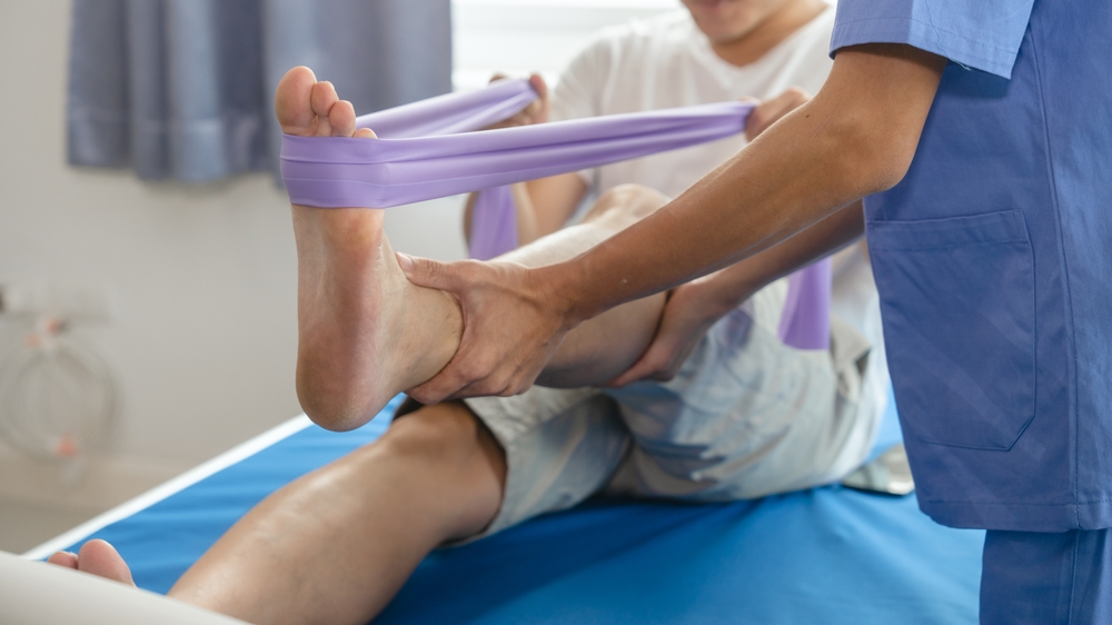
Conditions Treated
- Knee osteoarthritis and chronic knee pain
- Shoulder pain related to tendinopathy or bursitis
- Tennis elbow and golfer’s elbow
- Plantar fasciitis and chronic heel pain
- Achilles tendinopathy
- Facet joint–related back pain
- Nerve root–related radicular pain (e.g. sciatica)
- Osteoid osteoma and some other benign bone lesions
- Other chronic joint or tendon pain not responding to conservative treatment
* Suitability is always assessed individually after reviewing the clinical history and imaging.
Musculoskeletal & Sports Injury Interventions
Vertebroplasty
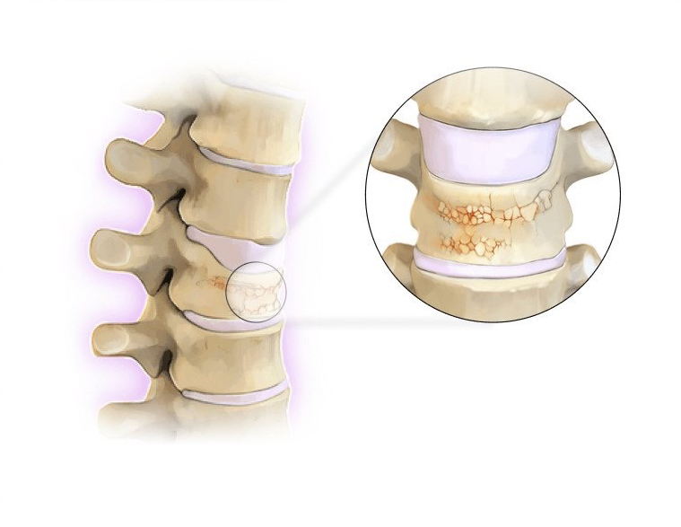
Vertebroplasty is a minimally invasive treatment used to alleviate severe spinal pain caused by new vertebral fractures, often resulting from osteoporosis, a condition where bone density is reduced.
During the procedure, a medical-grade acrylic cement is injected into the fractured vertebra using a needle guided by high-quality radiology imaging. The cement stabilises the bone, preventing further collapse and reducing the pain associated with fracture instability.
An interventional radiologist performs this procedure, providing an effective alternative to traditional surgical treatments for spinal fractures.
Your doctor may recommend vertebroplasty as part of your osteoporosis treatment if you suffer from severe pain due to a recent vertebral fracture, especially if the fracture occurred within the last three weeks.
This treatment is suitable for both hospitalised patients and outpatients who are struggling to manage their pain at home. The procedure is most effective when performed soon after the fracture, as early intervention can better prevent further vertebral collapse and enhance the benefits to bone health and pain relief.
Vertebroplasty is primarily used for osteoporotic fractures but may also be considered for fractures caused by conditions such as multiple myeloma or metastatic tumours.
Sacroplasty
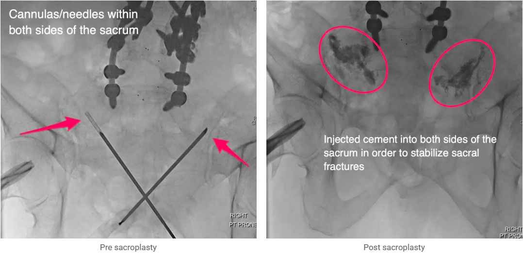
Sacroplasty is a specialized procedure designed to treat fractures of the sacrum, the large triangular bone at the base of the spine. These fractures are often due to osteoporosis, a condition where bones become weak and brittle, making them more susceptible to breaks. Sacral fractures can cause intense pain and significantly impact a patient’s mobility and quality of life. Sacroplasty offers a minimally invasive solution that helps stabilize the fracture and provides substantial pain relief.
During sacroplasty, a healthcare provider uses imaging guidance, such as live CT scans, or fluoroscopy, to precisely insert two hollow needles into the sacrum, one on each side of the fracture. A medical-grade bone cement is then injected into the fractured area through the needles. This cement hardens quickly, acting like an internal cast that stabilizes the bone and reduces pain. The entire procedure is performed under local anesthesia, typically taking less than an hour. Sacroplasty is performed on an outpatient basis, allowing patients to go home the same day and often resume normal activities shortly afterward.
This procedure is particularly beneficial for patients who have not responded to conservative treatments such as bed rest, bracing, or pain medication, or for those who experience significant side effects from these treatments. By directly targeting the fracture site, sacroplasty provides a quick and effective way to relieve pain and improve stability, helping patients regain their mobility and quality of life.
Sacroplasty is primarily used to treat sacral fractures caused by:
- Osteoporosis, particularly in elderly patients or post-menopausal women
- Long-term steroid use that weakens bones
- Trauma or other conditions that lead to sacral bone weakening
The procedure is ideal for patients who have not found relief through non-surgical treatments and are seeking a minimally invasive solution to manage their pain and stabilize their fractures.
Genicular Artery Embolisation (GAE)
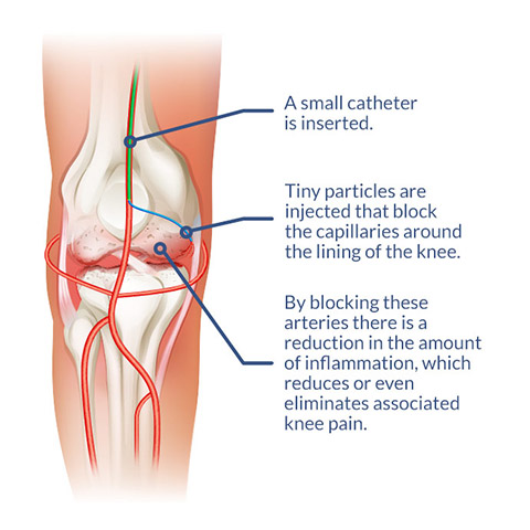
Genicular Artery Embolisation (GAE) is a modern knee pain treatment solution performed by Interventional Radiologists on individuals dealing with persistent and stubborn knee pain due to arthritis. This minimally invasive procedure is a modern option for those who have exhausted traditional remedies for their chronic knee pain.
Chronic knee pain is caused by an array of reasons, ranging from degenerative conditions like osteoarthritis to inflammatory disorders such as rheumatoid arthritis. Osteoarthritis is the most common cause and frequently leaves people dealing with pain that disrupts their daily lives and impedes their mobility.
Genicular Artery Embolisation is a minimally invasive procedure designed to alleviate chronic knee pain by targeting the lining of the joint which is normally thickened and inflamed by arthritis. By selectively reducing the blood flow to this joint lining, the thickening and inflammation in the joint is reduced, resulting in a reduction in pain.
During a GAE procedure, arteries are catheterised with a tiny tube and microscopic embolic agents (called microparticles) are injected to reduce blood flow to the painful regions of the knee. The overall effect is a reduction in pain, resulting in an improvement of function.
You may be referred for GAE if you have a history of chronic knee pain which is caused by knee arthritis and not due to an acute knee injury. Your eligibility for the procedure will be determined through a thorough medical evaluation and imaging studies, such as an Xray or an MRI, to ascertain the underlying causes of your knee pain. Not all knee pain can be solved by GAE, so it is important to have a complete diagnosis of the knee to better evaluate the potential for success. You may be referred to an orthopaedic surgeon if appropriate, during this assessment to ensure there is no current role for surgery for you.
Injections for shoulder pain
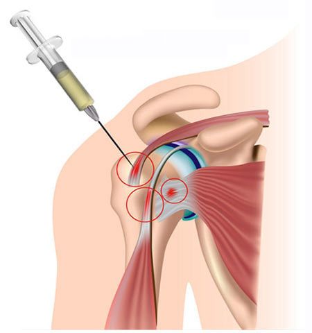
Osteoarthritis of the glenohumeral joint (ball and socket joint of the shoulder)
The shoulder is made up of two joints, The glenohumeral joint (the shoulder joint proper) and the acromioclavicular joint (sitting at the tip of your shoulder). Both joints can be affected by osteoarthritis. Osteoarthritis is known as a progressive degenerative condition which affects joints. This condition mostly affects people from their 50s onwards. Osteoarthritis develops when a joint is subjected under excessive pressure over a prolonged period of time. This increased joint pressure can result in the joint surfaces rubbing together which, in turn, causes the articular cartilage (shock absorbing inner layer of the joint) to wear. If the joint is continually stressed the articular cartilage wears away leaving the underlaying bone surfaces to become exposed. Joint irritation can result in the inner lining of the joint capsule (called the synovial membrane) to become irritated and inflamed. This is called (known as synovitis) and is often the cause of the pain associated with osteoarthritis.
General signs and symptoms associated with osteoarthritis of the shoulder include:
- Pain surrounding the shoulder joint. This can be hard to locate.
- Shoulder joint stiffness (especially first thing in the morning).
- Reduced range of motion. Some people find difficulty with raising the hands above their head.
- Change in shape. An osteoarthritic joint can appear larger and thickened as the disease process progresses.
Frozen shoulder
A frozen shoulder is an incredibly common condition which causes the shoulder joint to become stiff and extremely painful. Symptoms often start without any warning, frequently without incident or accident. Frozen shoulders typically affect people in their 40’s to 60’s and most commonly affects women and Diabetics. The pain and stiffness associated with a frozen shoulder can last for a few years and so it is paramount that a frozen shoulder is treated correctly as soon as possible.
General signs and symptoms associated with frozen shoulder are:
- Significant pain
- Increasing stiffness in the shoulder joint. Tasks such as dressing and washing your hair becomes challenging.
- Pain that wakes you during the night
Research states that the most effective treatment available for frozen shoulder is injection therapy. After an injection pain levels can be significantly reduced allowing you to partake in a period of intensive physiotherapy, aimed at increasing the flexibility and function in the arm. Follow this link to find out more about frozen shoulder.
Rotator cuff pain
The rotator cuff is a series of important muscles who’s tendons attach around the shoulder joint. The muscles (and most importantly, their tendons, are supraspinatus, infraspinatus, subscapularis and teres minor). The rotator cuff contracts together to hold the shoulder joint in position during movement and so are pivotal in producing pain free movement. The rotator cuff is susceptible to injury. Injury can occur due to repetitive movements undertaken during sports and hobbies, after an accident or fall or can slowly develop over time, seemly without any reason.
Rotator cuff related pain encompasses a series of pathologies which can affect the rotator cuff tendons of the shoulder. These include tendinitis (an inflamed and irritated tendon), tendinopathy (wear and tear of a tendon) or tear (tears can be partial, leaving a portion of the tendon unaffected or complete, involving the whole of the tendon).
General signs and symptoms associated with rotator cuff pain are:
- Pain radiating from the shoulder down the outside of the arm towards the elbow and around the back of the shoulder and shoulder blade.
- Pain which is made worse when lifting the arm above the head or out to the side.
- A sensation of weakness. This is mostly evident when trying to bring your arm back down from an overhead activity.
- Pain that wakes you at night
When conservative management including physiotherapy and rehabilitation exercises have not been effective then an injection can be very useful to reduce pain and allow you to rehabilitate the rotator cuff tendons.
Subacromial bursitis
A bursa is a fluid filled sack which provides a friction free surface for different structures to slide past each other allowing pain free during movement to occur. There are around 150 bursae found throughout the body. The shoulder is a highly mobile joint, capable of a wide variety of movements and so has many bursae surrounding it. The largest, and by far the most pain sensitive, is the subacromial bursa. The subacromial bursa sits above the rotator cuff tendons and provides a friction free surface for shoulder motion to occur. A bursitis occurs when a bursa becomes irritated and inflamed. This can occur due to a single incident or accident but most commonly happens gradually due to low level, repetitive movements.
General signs and symptoms associated with rotator cuff pain are very similar to rotator cuff related pain and include:
- Pain radiating from the shoulder down the outside of the arm towards the elbow and around the back of the shoulder and shoulder blade.
- Pain which is made worse when lifting the arm above the head or out to the side.
- A sensation of weakness. This is mostly evident when trying to bring your arm back down from an overhead activity.
- Pain that wakes you at night
This is a highly painful condition and is an incredibly common condition treated with both physiotherapy and injection therapy. When physiotherapy and rehabilitation exercises have not worked then an injection can be considered.
Calcific tendinopathy
Calcific tendinitis of the shoulder is an incredibly painful condition which has a rapid onset of significant pain which often radiates from the shoulder down the arm. Many patients even report attending A&E due to the severity and rapidity of their symptoms. All activities of daily living are affected and sleep can be challenging. A calcific tendinitis can occur throughout the rotator cuff of the shoulder but is most common in the supraspinatus tendon. It is still not fully understood why some people develop a build-up of calcium within a tendon however it is believed to be associated with repetitive tasks. Being female and in the 4th and 5th decade of life are also common sufferers.
General signs and symptoms associated with calcific tendinitis are:
- A sudden and significant onset of severe pain in the shoulder and upper arm.
- A significant reduction in the ability to use the arm (due to pain).
- Pain that is worse first thing in the morning.
- Pain that wakes you at night.
Biceps tendinitis
Biceps tendinitis typically causes pain located at the front and side of the shoulder. Although it can affect anyone at any age it is most commonly seen in younger sporty patients. Biceps tendinopathy occurs due to repetitive movements and is often associated with manual jobs such as building or sports such as tennis. When the biceps tendon is subjected to prolonged periods of stress it can become irritated and inflamed, this is known as tendinitis. If the tendon continues to be subjected to repeated episodes of tendinitis the tendon structure can become compromised resulting in a weakened tendon. This is called tendinopathy. Furthermore, the biceps tendon can rupture however, the bicep tendon can easily be assessed using diagnostic ultrasound and the most effective treatment selected. If physiotherapy and a specific rehabilitation program does not resolve your symptoms then a corticosteroid injection may be required.
General signs and symptoms associated with a biceps tendinitis are:
- Pain both at the front and side of the shoulder.
- Pain when lifting the arm above the head, especially out in front of you.
- Tenderness located at the front of the shoulder.
Acromioclavicular joint sprain
The acromioclavicular joint is located at the tip of the shoulder (the junction between the acromion of the scapular and the clavicle or collarbone). This joint is highly susceptible to injury during contact sports such as a tackle during a rugby match or after falling from a bicycle. Furthermore; the acromioclavicular joint is commonly affected by osteoarthritis.
General signs and symptoms associated with a acromioclavicular joint sprain are:
- Focal pain located at the top of the shoulder.
- Pain on palpation of the joint itself.
- Pain lying on your affected side.
- Pain when moving your arm across your body.
Nerve root injection for Back pain and sciatica
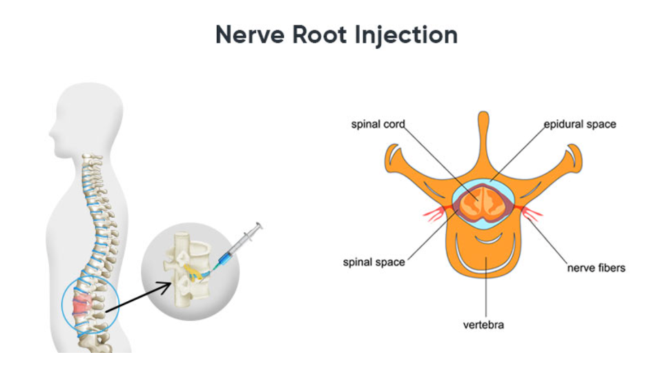
A nerve root injection is a very common procedure where a mixture of local anaesthetic and steroid (anti-inflammatory) is injected around a swollen or compressed spinal nerve, to help relieve the pain and inflammation of nerve pain.
Nerve pain and inflammation is most commonly due to compression from a disc herniation (disc prolapse) or stenosis. This is typically due to disc degeneration, or “wear and tear,” in the spine. Nerve root injections, also known as nerveblocks, are commonly performed in the lower back, or lumbar spine, to treat radiating leg pain, known as sciatica. Nerve root injections can also be performed in the neck (cervical spine) to treat symptoms of arm pain (brachialgia) or cervical radiculopathy.
If sciatica pain or nerve pain cannot be controlled with pain medications then a trial of a nerve root injection (steroid injection) is a very appropriate and highly recommended treatment option. It is a very safe procedure that may help improve painful symptoms. The procedure is typically performed under CT guidance by a radiologist, or by a pain physician under sedation with the aid of an x-ray.
Why does Dr Aser recommend a nerve root injection?
In the cascade of treatment for sciatica and other nerve root compressive problems, such as radiculopathy, following failure of medications to control the pain, a nerve root injection is a safe and simple treatment that may help relieve the symptoms of nerve pain.
The injection of local anaesthetic and steroid reduces the inflammation in the nerve to help with pain. In most patients with disc prolapse it is worthwhile trying a nerve root injection prior to consideration for surgery. If the nerve root injection helps control the pain, surgery may be avoided or delayed. In addition, if the nerve root injection does help, it is diagnostic that the compressed nerve is the symptomatic level, and is predictive of a good surgical response.
Procedures Offered
Image-Guided Joint Injections
Accurate delivery of medications (e.g. steroid, viscosupplement or other agents, as appropriate) into affected joints under ultrasound or fluoroscopic guidance.
Common joints include the knee, shoulder, hip, ankle and small joints of the foot and hand.
Aim: reduce inflammation and pain, improve mobility, and complement physiotherapy.
Nerve Root and Spinal Injections
Targeted injections around irritated nerve roots or facet joints under fluoroscopy or CT guidance.
Used for radicular pain (e.g. sciatica) and selected cases of chronic spine-related pain.
Aim: reduce nerve irritation, improve function and help guide further management.
PRP (Platelet-Rich Plasma) Injections
A small amount of the patient’s own blood is processed to concentrate platelets, then injected into the affected tendon or joint under ultrasound guidance.
PRP may support tissue healing in selected patients with tendinopathy or early joint degeneration.
Often used alongside structured rehabilitation programmes.
Osteoid Osteoma Treatment (Ablation)
Minimally invasive treatment of osteoid osteoma and some benign bone lesions using image-guided thermal ablation.
A tiny probe is placed into the lesion under CT guidance and heat is applied to destroy it.
Most patients experience rapid and often complete pain relief, usually as a day case.
MSK Embolization
Catheter-based procedure to treat chronic tendon or joint pain where abnormal tiny blood vessels contribute to inflammation and pain.
Through a pinhole puncture in the wrist or groin, a catheter is guided to the target area and selective embolization of abnormal vessels is performed, preserving normal circulation.
Can be an option when symptoms persist despite physiotherapy, medications and injections, or when surgery is not preferred.
What to Expect
Before the procedure
Consultation to review symptoms, imaging and previous treatments.
Additional imaging (e.g. MRI, ultrasound, CT) may be arranged if needed.
Clear instructions will be provided regarding fasting, medications and the day of the procedure.
During the procedure
Most procedures are done with local anaesthesia; light sedation can be used if required.
Ultrasound, fluoroscopy or CT guidance is used for accurate needle or catheter placement.
Many procedures take between 20–90 minutes, depending on complexity.
After the procedure
Short observation period in the day-care unit.
Mild soreness at the injection or puncture site is common and usually settles quickly.
Specific advice will be given on activity, physiotherapy and follow-up.
Pain improvement can be immediate (especially after some injections) or gradual over weeks to months, particularly with PRP and embolization.
Why Choose Dr Aser for MSK and Sports Pain Procedures?
- Subspecialty training in Musculoskeletal Radiology and Interventional Radiology in the UK and Canada
- Experience across a wide spectrum of MSK interventions, not just a single technique
- Access to advanced imaging and Angio Suite facilities in a tertiary centre
- Collaboration with orthopaedics, sports medicine, rheumatology, physiotherapy and pain specialists
- Focus on clear explanation, realistic expectations and only recommending procedures where there is a reasonable chance of benefit
FAQ
1. How long does relief from MSK Embolization last?
Relief after MSK embolization is durable. In many patients, improvement begins within 2–4 weeks and can last more than 2 years.
2. Is MSK Embolization safe?
Yes. MSK Embolization is performed by experienced vascular specialists in a certified ambulatory surgery center. Risks are low, and complications are uncommon.
3. Does MSK Embolization help arthritis?
MSK Embolization may provide relief for patients with arthritis by reducing abnormal blood flow and inflammation that contribute to joint pain. For knee arthritis, the procedure is often referred to as Genicular Artery Embolization (GAE). Patients with arthritis in other joints may also benefit, depending on the severity and location of their pain.