Overview
We offer advanced, image-guided treatments for liver tumors, biliary obstruction, and a wide range of oncological and hepatobiliary conditions. Using CT, ultrasound, and fluoroscopic guidance, our specialists deliver precise, targeted therapies such as tumor ablation, chemoembolization, radioembolization, and biliary drainage or stenting. These minimally invasive procedures help control cancer, relieve symptoms, improve liver function, and often serve as effective alternatives to major surgery—enhancing both comfort and quality of life for patients.
Advantages
- Advanced Portal Hypertension Management: TIPS provides an effective, minimally invasive solution to reduce portal pressure, preventing life-threatening variceal bleeding and managing refractory ascites.
- Targeted Biliary Relief: PTBD ensures precise drainage of bile in cases of obstruction, jaundice, or malignancy, reducing infection risk and improving liver function.
- Quick Symptom Relief: Ascitic and pleural fluid aspiration delivers immediate comfort by removing excess fluid, addressing breathing difficulty, abdominal discomfort, or diagnostic needs.
- Minimally Invasive Techniques: All procedures use imaging guidance and percutaneous approaches, reducing surgical trauma, recovery time, and hospital stay.
- Customized Patient Care: Treatment plans are based on individual liver health, disease severity, and underlying conditions, ensuring safety and optimal outcomes.
- Rapid Monitoring and Follow-Up: Doppler, imaging, and lab assessments enable real-time evaluation of procedure success and early detection of complications.
Treatments
Biliary Drainage And Stenting

Biliary drainage is the insertion of a tube into the bile duct. This is mainly carried out when the bile ducts are blocked.
You will be referred for biliary drainage and stenting because of gallstones impacted in the ducts, Injury to or narrowing in the bile ducts from previous surgery and involvement of cancer in the ducts.
Cryoablation for Kidney Tumours

Cryoablation is an interventional radiology procedure that uses extreme cold to destroy cancerous kidney tissue.
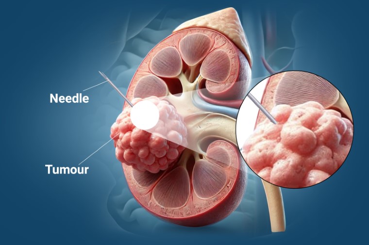
During the procedure, a special needle (cryoprobe) is inserted into the tumour under image guidance. The cryoprobe freezes the tumour tissue by circulating a freezing agent, typically argon gas, to create extremely cold temperatures. The freezing process destroys the cancer cells, and as the tissue thaws, the damaged tumour cells are gradually reabsorbed by the body.
Cryoablation is a minimally invasive alternative to surgery, offering effective treatment with less downtime and faster recovery.
Your doctor may refer you for cryoablation if you have a kidney tumour that is small or localised and if surgery is not the best option for you. Cryoablation is particularly beneficial for patients who may not be ideal candidates for surgery due to age, health conditions, or personal preference.
This procedure is often used for patients with early-stage kidney tumours, including those with renal cell carcinoma (the most common type of kidney cancer), or for patients with other small renal masses. Cryoablation is designed to treat the tumour while sparing as much of the healthy kidney tissue as possible.
Image Guided Tumour Ablation

Tumour ablation is the process whereby a needle is placed directly into a tumour and a treatment is delivered aiming to kill the tumour in place. This can be achieved through a variety of ways but the most common is via heating or cooling the tumour. Other approaches that can also be used in difficult tumour locations include injection of a chemical (usually alcohol or acetic acid) or use of electricity passed through the cells.
If you have been diagnosed as having a tumour, image guided tumour ablation is used as an alternative to surgery with curative intent if surgery is felt to be too high risk or for palliation (improving quality of life or length of life, but not destroying all tumours).
Occasionally, ablation may be the best treatment due to it being less invasive than surgery with similar efficacy, such as in some benign tumours of the bone or in small primary liver cancers.
Selective Internal Radiation Therapy (SIRT)

SIRT, also known as radioembolisation, is a nuclear medicine treatment designed for primary and metastatic liver cancers like hepatocellular carcinoma (HCC).
During the procedure, tiny radioactive microspheres are injected directly into the liver arteries that are supplying the tumours. These microspheres concentrate in the tumours and release radiation over a period of weeks, effectively shrinking or destroying the cancerous cells while minimising damage to surrounding healthy tissue.
SIRT is a vascular interventional radiology procedure often recommended for patients whose liver tumours cannot be surgically removed or treated with other methods.
Selective Internal Radiation Therapy is a minimally-invasive interventional radiology treatment using implanted radiation that is often recommended when surgery or other ablative treatments are not suitable for addressing liver tumours.
If you have been diagnosed with liver cancer like Hepatocellular carcinoma (HCC) or liver metastases from another cancer (such as colorectal cancer or breast cancer), SIRT might be an option. However, it’s important to note that SIRT is not suitable for all patients, especially those with severe liver failure. Your doctor will assess your liver function and overall health to determine if SIRT is appropriate for you.
Transarterial Chemoembolisation For Liver Cancer (TACE)

TACE is a minimally invasive procedure in which chemotherapy drugs are delivered directly into the liver tumours through the liver arteries using interventional radiology, helping to destroy cancer cells while sparing healthy tissue.
The term “transarterial” refers to delivering the treatment through the arteries, and “chemoembolisation” involves using both chemotherapy drugs and an embolic agent to block the blood supply to the tumour. By cutting off the tumour’s blood supply from the hepatic artery, the chemotherapy remains concentrated in the liver, making it more effective against the cancer cells while reducing side effects on the rest of the body.
TACE is typically recommended for patients with liver cancer whose tumours cannot be removed surgically or treated with ablation (destroying tissue using heat or cold). It is commonly used for those with primary liver cancer (hepatocellular carcinoma) or liver metastases (cancer that has spread to the liver from another part of the body). TACE can slow the growth of tumours, improve quality of life, and extend survival.
Percutaneous Image Guided Biopsy

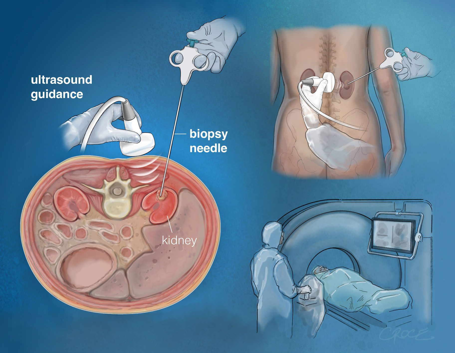
A biopsy is a method of using a special needle to take a small piece of tissue from inside the body for analysis in the laboratory, normally to diagnose or exclude cancer. This could be from an organ such as the liver, kidney or from a lymph node or other area such as the lining of the abdomen (peritoneum).
The biopsy needle is advanced through a small cut in the skin overlying the area to be biopsied and image guidance is used to plan a path into the target. The sample of tissue is then sent to a laboratory for examination under the microscope.
The reason for requesting this test is that your referring doctor seeks an assessment of the tissue to look for any abnormality or disease that may be present. Often this is used to exclude or diagnose cancer or infection and to obtain specific information about the cells involved.
Transjugular Intrahepatic Portosystemic Shunt (TIPS)

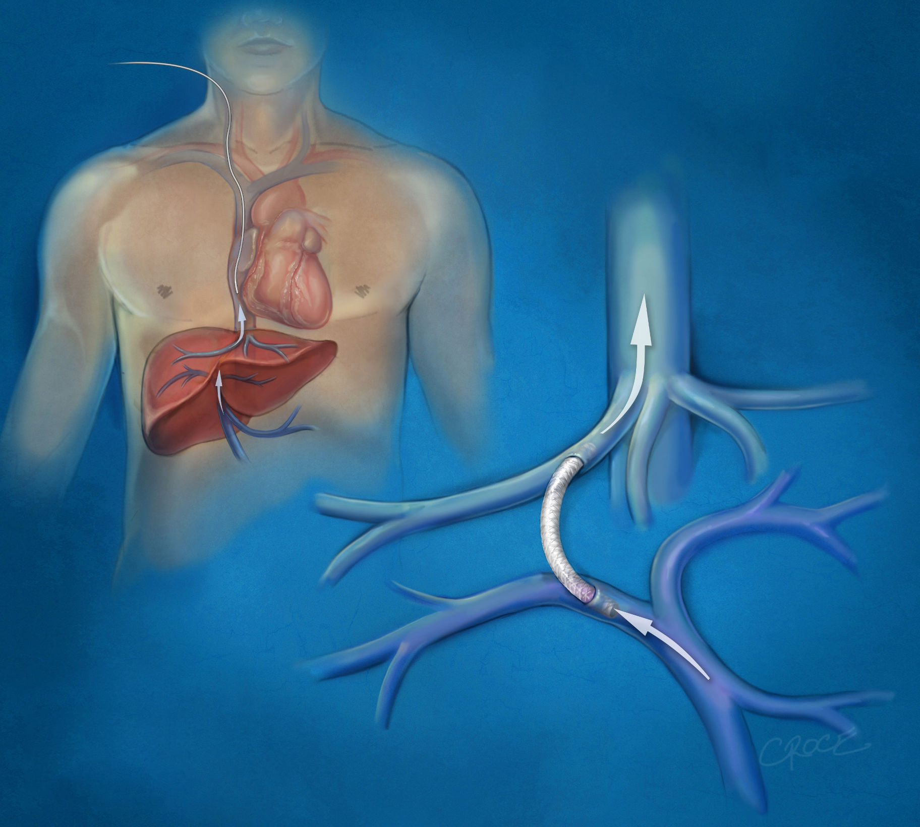
TIPS is a procedure that lowers pressure in the portal vein. That’s the vein that moves blood to your liver.
The TIPS procedure is usually performed in patients with liver cirrhosis. If you have this condition, the normal blood flow through your liver is blocked by scar tissue, which increases the pressure in your portal vein. The increased pressure in your portal vein makes thin veins in your stomach become abnormally enlarged and so are at risk of bleeding. Another symptom of liver cirrhosis is an abnormal collection of fluid (ascites) in the abdominal cavity.
You may be advised to undergo TIPS if you have varices (dilated veins caused by high portal vein pressure) which bleed acutely or recurrently and have not responded to other treatments.
FAQ
1. Are these procedures minimally invasive?
Yes. All treatments, including ablation, chemoembolization (TACE), radioembolization (Y90), biliary stenting, and TIPS, are performed using image guidance through tiny skin punctures, without open surgery.
2. Which imaging techniques are used during these procedures?
Depending on the treatment, specialists may use:
- Ultrasound
- CT (Computed Tomography)
- Fluoroscopy (real-time X-ray)
These ensure high accuracy and safety.
3. Do I need general anesthesia?
Most procedures require only local anesthesia and mild sedation, keeping patients comfortable while avoiding the risks of general anesthesia. More complex interventions may use deeper sedation if needed.